
Back pain is one of the most common reasons people end up in a doctor’s office, and one of the most common reasons they get an MRI ordered. The honest part is that most back pain doesn’t need an MRI right away. The useful part is that when it does, the scan answers real questions. Here’s how to tell the difference.
Most Back Pain Resolves on Its Own
The vast majority of low back pain gets better with time and movement, regardless of treatment. That’s why most guidelines recommend against imaging in the first several weeks unless there’s something specific going on. A lumbar MRI shows anatomy — discs, nerves, the spinal canal. It doesn’t show “pain.” Lots of people with bulging discs have no symptoms at all, and lots of people with symptoms have unremarkable scans. That’s not a reason to avoid imaging; it’s a reason to use it when it’s actually indicated.
When Imaging Makes Sense
Your doctor is more likely to order a lumbar spine MRI when:
- Back pain radiates below the knee — sciatica — or you have numbness or weakness in a leg.
- The pain has lasted more than about six weeks despite conservative treatment.
- There’s a sudden onset of back pain with a fever, or urinary or fecal incontinence — these are urgent red flags.
- You had significant trauma to the spine.
- A spine surgeon needs imaging for pre-surgical planning.
If your symptoms fit one of those, an MRI is genuinely useful. If not, waiting and treating conservatively is usually the right call, and most clinicians will tell you the same.
What a Lumbar MRI Shows
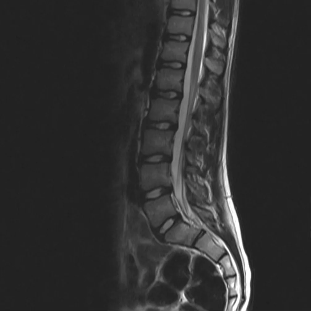
The scan images the five lumbar vertebrae (L1–L5), the discs between them, the spinal canal, and the nerve roots. A radiologist reads the images for:
- Disc herniation or bulge, including which nerve root is being compressed — the thing that explains sciatica.
- Spinal stenosis, a narrowing of the spinal canal that can cause pain with standing and walking.
- Nerve compression causing radiating pain.
- Degenerative disc disease and related wear.
- Fractures, inflammation, or other changes that X-ray can’t characterize.
The full breakdown of what the study covers and how to prepare is in our earlier post on the lumbar spine MRI procedure.
What It Costs and What the Visit Is Like
A lumbar MRI at Sunset Hills Imaging is a flat $399 — the scan, the radiologist’s read, and a CD of your images, with no facility fee and no separate bill. It’s also one of the easier scans for anxious patients: you go in feet first, and your head stays outside the scanner for most of the study. The scan takes about 25 minutes, the whole visit runs about an hour, and you’re free to leave as soon as it’s done.
What If the Scan Comes Back Unremarkable?
If your lumbar MRI comes back unremarkable, that’s not a wasted scan — it’s information. It tells you and your doctor that the pain isn’t coming from a disc, a nerve, or a structural problem in the spine, which redirects the search toward muscles, movement patterns, or other sources. An unremarkable report is a step forward in a workup, not a dead end. The same logic cuts the other way: a report with findings doesn’t automatically mean those findings are causing your pain, and your provider is the right person to connect the images to your symptoms.
The pricing page shows the full comparison against hospital billing. If you have a doctor’s order and want to get the scan done, call (314) 834-7226 — same-day appointments are often available.