
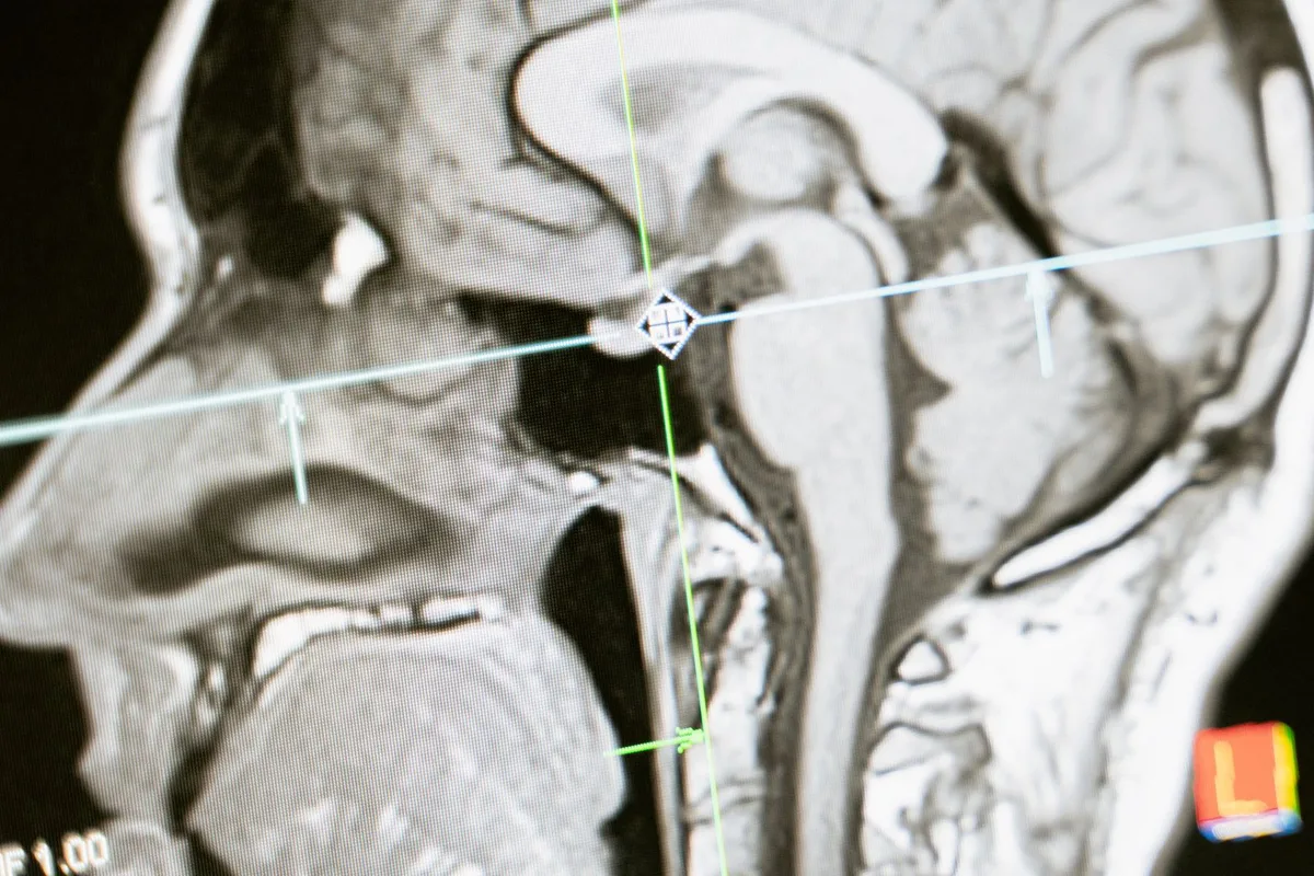
A brain MRI is different from most other scans in one important way: your head goes inside the machine. That’s the thing patients want to know about before they arrive, so we’ll be direct about it here, along with everything else you can expect from the appointment.
Why It’s Ordered
Non-contrast brain MRI is the standard study for persistent headaches that changed in pattern or severity, dizziness, memory concerns, vision changes, and a range of neurological symptoms. It can show brain tissue, the ventricles, and white matter in far more detail than a CT scan, and it uses no radiation. If your doctor ordered this study, they’re looking for structural answers — and an unremarkable report is useful information too.
No IV, No Contrast
One thing that surprises people: at our clinic, brain MRIs are non-contrast. There’s no IV, no injection, and no dye. For many clinical questions — headache workups, white matter changes, structural evaluation — contrast simply isn’t needed. If your order genuinely requires contrast for a question like tumor characterization or active MS lesion dating, we’ll tell you honestly and point you to a facility that administers it rather than run an incomplete study.
The Head Coil and the Scan
For a brain MRI, you lie on your back with a padded coil around your head. Your head is inside the scanner for the full study — that’s unavoidable with a brain scan. The scan itself takes about 30 minutes, and the whole visit runs about an hour including check-in and positioning.
A few practical notes so nothing surprises you:
- The machine is loud — expect banging and buzzing. You’ll wear earplugs and over-ear hearing protection.
- Skip hairspray, gel, and dry shampoo the day of the scan, and remove earrings and facial jewelry. Some eye makeup contains metallic pigment, so leave it off.
- You can talk to your technologist through speakers at any time, and you’ll have a call button.
- We’ve scanned thousands of brain studies, and the honest truth is that most people settle in once the first sequence starts. The banging is the machine working — it’s not a problem with the scan, and the technologist can hear you the whole time.
What the Report Can and Can’t Tell You
A brain MRI shows structure, and the radiologist reads it for structural findings: white matter changes, old or chronic infarcts, lesions, ventricular size, and conditions like hydrocephalus or Chiari malformation. What it can’t do is explain every symptom you’ve ever had. Headaches, dizziness, and memory concerns often come back with imaging that looks completely normal — and that normal result is still valuable, because it rules out the structural causes and lets your provider focus on the rest of the workup. The scan is one piece of the picture, and the report is written for your doctor to fit into the rest of it.
What Happens After
When the scan is done, the table slides out and you’re free to go — no recovery time, since there’s no contrast. A board-certified radiologist reads the images, and the report goes to your ordering provider within 24–48 hours. Ask us and we’ll email you a password-protected copy, and you’ll get a CD of your images.
The complete walkthrough of how to prepare, what to wear, and what happens during any MRI is in our what to expect guide. If you’d rather talk it through first, call us at (314) 834-7226 — especially if you’re nervous about your head being in the machine. We’re used to first-timers, and we’ll walk you through exactly what your study involves.